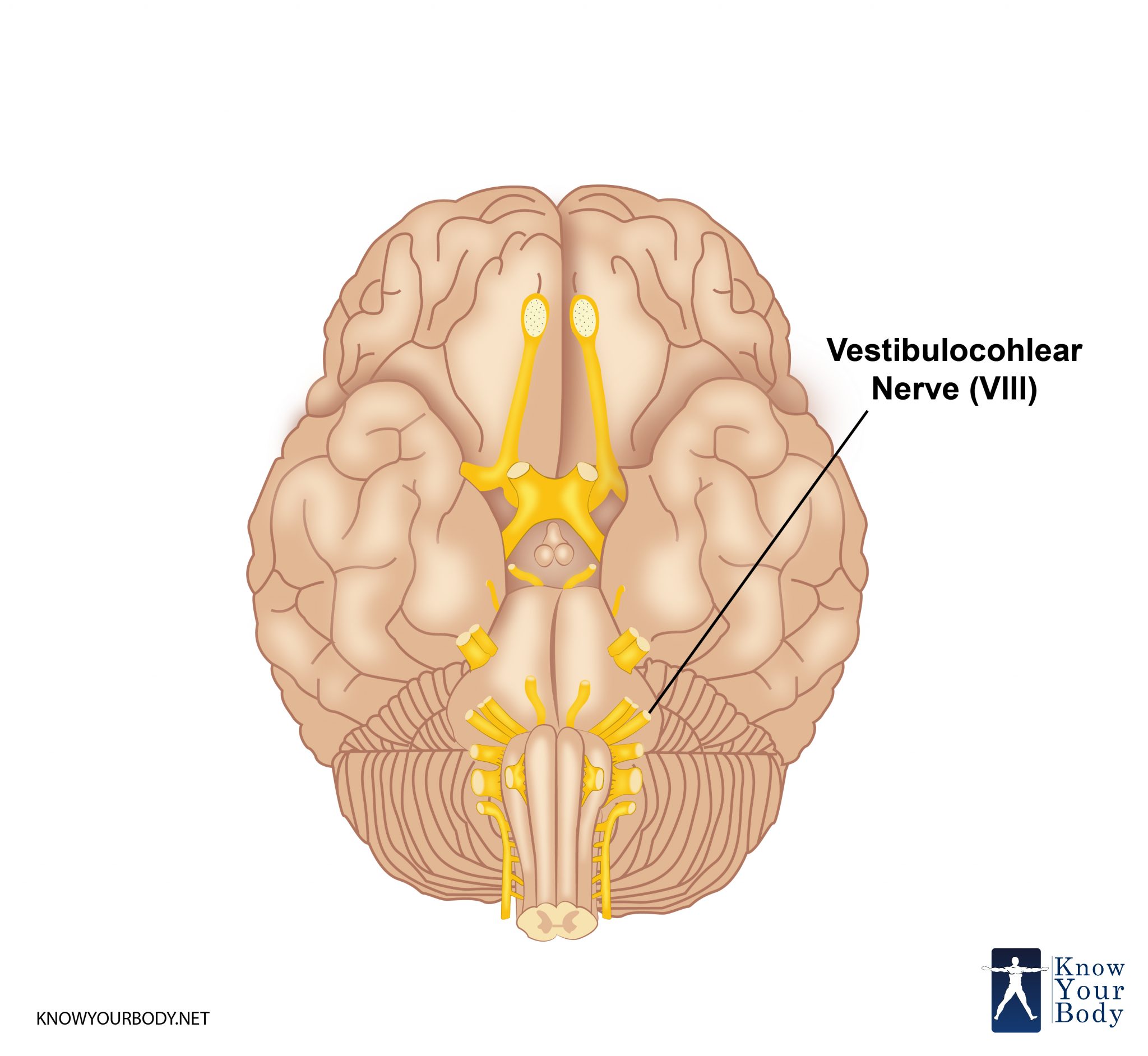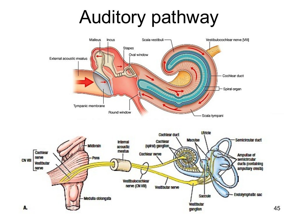

Via a complex circuit of auditory nerve pathways to the auditory cortex and other Once sound is converted to electrical signals in the cochlea, these signals travel Step 4: Your brain interprets the signal. Blasting hair cells with noise is akin to trees in a hurricane, struggling to remain standing. They’re also quite fragile: Loud sounds can damage or even destroy them, and once they’re destroyed, they can’t be repaired-and you’ll feel the effects of noise-induced hearing loss. Note: Hair cells play a vital role in your hearing. These hair cells translate the vibrations from sound waves into electrical impulses that then travel along a complex pathway of nerve fibers to the brain. You’re born with about 16,000 of these hair cells, according to the Centers for Disease Control and Prevention (CDC). (While less common, some people have low-frequency hearing loss or mid-range hearing loss.)

This means the hair cells responsible for detecting high frequencies are damaged. For example, many people with hearing loss have high-frequency hearing loss, making it harder to hear high-pitched sounds. The tiny hair cells lining the cochlea are stimulated by different frequencies. In the organ of Corti, vibrations are finally transformed into electrical energy by cells known as hair cells (stereocilia). Vibrations from the stapes push on the oval window, and set up pressure waves in the fluid-filled cochlea, the snail-shaped inner ear that contains the organ of Corti. Step 3: Sound moves through the inner ear (the cochlea) The orientation of the three bones allows them to function as a lever, amplifying the sound energy as it moves from the relatively large tympanic membrane to the relatively small oval window. This last bone-the stapes-is connected to the oval window, which is a membrane separating the middle ear from the inner ear. The shapes of the ossicles provide inspiration for their nicknames.

The incus, in turn, is attached to the stapes (the “stirrup” or “footplate”). The bone directly attached to the eardrum is the malleus (“the hammer”), which is connected at its other end to the incus (“the anvil”). These three bones are the smallest ones in your body. When the eardrum vibrates in response to sound waves, these bones are set into motion as well. Here’s how that process unfurls: The eardrum is attached to a chain of three small bones, known as the ossicles. In this part of the ear's anatomy, sound waves are amplified before they are delivered to the inner ear. Step 2: Sound moves through the middle earīehind the eardrum is the middle ear. “The eardrum is a paper-thin layer of a membrane that essentially vibrates as soon as sound waves hit it-very similar to a drum,” Dr. Next, sound waves hit the eardrum, or tympanic membrane, setting it in motion. The pinna is the visible portion of your ear, and its funnel-like shape is well-engineered: As sound hits the pinna, it filters and amplifies sound waves, and chutes them along into the ear canal, Dr. When a sound occurs, it enters the outer ear, also referred to as the pinna or auricle. How humans hear Step 1: Sound waves enter the ear. Our brain uses these signals to organize and communicate with the external world. “It's quite a complex and intricate system,” Omid Mehdizadeh, MD, an otolaryngologist ( ENT) at Providence Saint John’s Health Center in Santa Monica, Calif., tells Healthy Hearing.īut how exactly does this process unfold? We've put together a step-by-step explanation of how people hear-from the moment sound waves arrive to the outer ear, then travel through the middle and inner ear and transform into meaningful signals sent on to the brain.

Pause to think about it, and our body’s ability to translate noise into sound is incredible.


 0 kommentar(er)
